Engineering approaches grounded in immunology hold the key to the discovery and development of novel treatments for cancer, infectious disease, and autoimmunity. To this end, the overarching goal of our laboratory is to engineer immunity through a fusion of immunology with biotechnology and materials chemistry, employing a materials science-centric approach to create new therapies based on the controlled modulation of the immune system. more >>
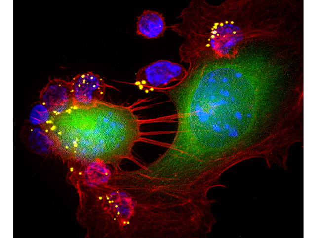
Licensed to kill. This image shows killing in action. The B16F10 tumor cell (green, CFSE) on the left attacked by multiple T cells (blue, DAPI) carrying nanopartilces (yellow, DiD) shows morphological alterations such as rounding, characteristic of poor cell health, while the tumor cell on right with T cell conjugates just being formed, is healthy. Entry is 100X. Image by Sudha Kumari/Yiran Zheng.
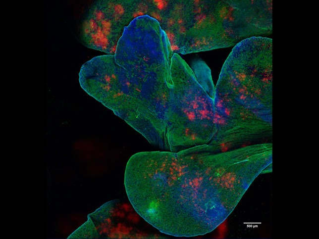
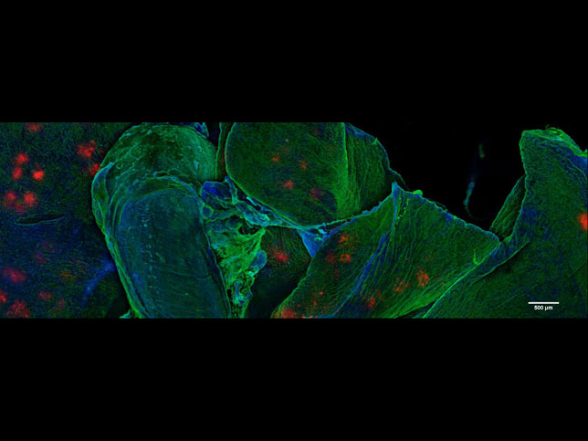
Polymer drug depots in the lungs. Intratracheal delivery was used to deposit polymer microparticle drug depots in the lungs of mice. Shown are multiple lobes of lung with many particle depots. Green: tomato lectin (endothelial staining); cyan: nuclei (DAPI); red: microparticles (DiD). By Greg Szeto/Adelaide Tovar
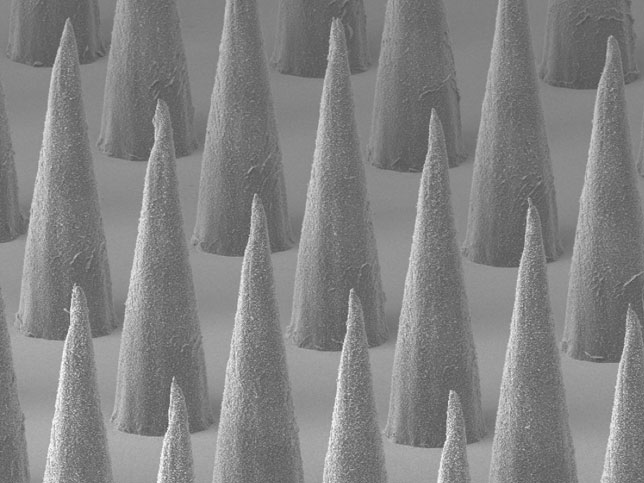
Microneedles
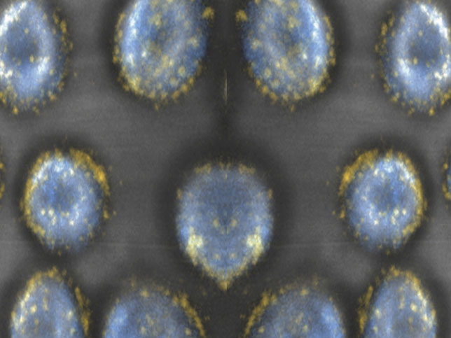
Pharmacytes montage
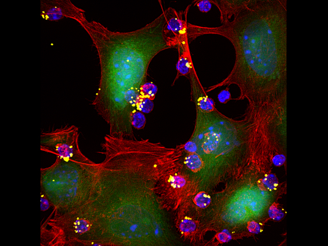
Lethal encounter.The image indicates robustness of immunosurveillance, where fewer T cells are able to find the tumor cells and form conjugates- the first and key step towards a killing event. This image is a larger view showing many T cell-tumor cell conjugate formation events occurring simultaneously. B16F10 tumor cell (green, CFSE) T cells (blue, DAPI); nanopartilces (yellow, DiD); phalloidin, Alexa568.Entry is 60X. Image by Sudha Kumari/Yiran Zheng.
Polymer drug depots in the lungs and trachea. Intratracheal delivery was used to deposit polymer microparticle drug depots in the lungs of mice. The trachea is shown near the center of the image while lobes of the lung are shown behind/to the sides. Green: tomato lectin (endothelial staining); cyan: nuclei (DAPI); red: microparticles (DiD). By Greg Szeto/Adelaide Tovar
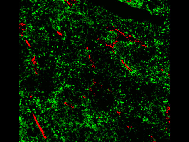
TA99: Confocal microscopy of a cryosection of B16F10 melanoma tumor and associated vascular endothelium, 10x magnification (green: Trp1 Alexa Fluor 488; red: CD31 PE). By Courtney Diamond/Gregory Szeto
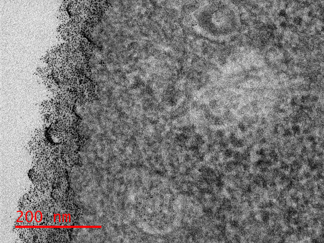
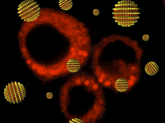
Membrane packing: In the Irvine lab, we are studying the interaction of amphiphilic gold nano particles (amph-NPs) and cell membrane interaction.Amph-NPs embed within lipid membranes and traverse into the cytosol subsequently. We investigate the effect of amph-NP's ligand structure and chemistry to better understand the role that governs the membrane penetration. This image shows short amphiphilic ligand protected amph-NPs labeling the glycocalyx of epithelium-derived cancer cell. By Yu-Sang Yang.
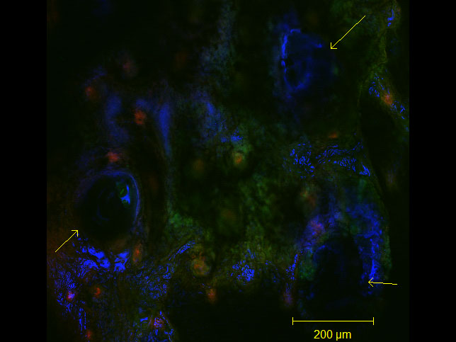
Delivery of sucrose/virus-tipped microneedles into the ears of transgenic mice. Yellow arrows indicate microneedle penetration pits/points. color key--blue: sucrose MN tips (Alexa Fluor 647), red: wild-type cells (tdTomato), green: virus infected cells/gene recombination (EGFP);  taken at 10X. By Lauren M Adelaar, Gregory L Szeto
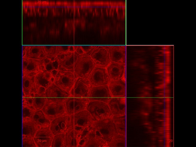
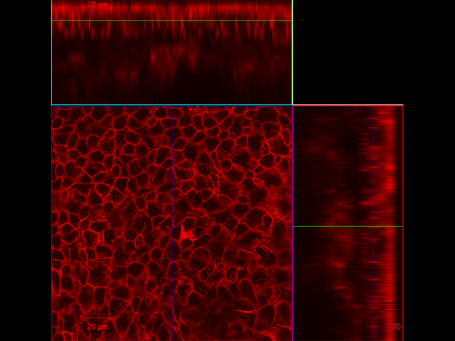
LMA2: Suprabasal layer within the epidermis of mtmg mouse ear (taken at 63x). red: tdTomato beta-actin. By Lauren M Adelaar, Gregory L Szeto
LMA3: Keratinocytes in epidermis of mtmg mouse (taken at 63x). red: tdTomato beta-actin. By Lauren M Adelaar, Gregory L Szeto
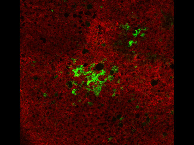
In-transit metastases in a lymph node from a spontaneously arising primary BRAFV600E PTEN-/- melanoma in mice. Red: wild-type cells; green: cancer cells 10x mag. By Lauren Adelaar, Gregory Szeto
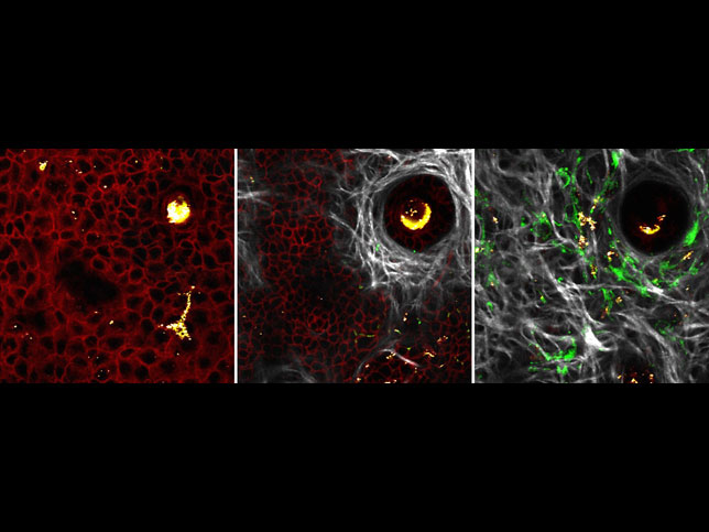
Layers of a melanoma. Microneedle-delivered tamoxifen induces a malignant melanoma in a genetically engineered mouse. Different depths of skin are shown with the invasive phenotype of melanoma cells being demonstrated. Red - normal cells; green - tumor cells; white - collagen. By Lauren Adelaar/Michael Zhang/Greg Szeto, Jeffrey Wyckoff
Striped nanoparticles
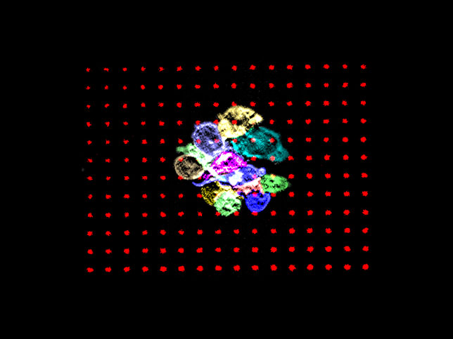
Aggregated T cells on agonist microdots: The microdot image is of aggregated T cells on agonist microdots. It shows the activation induced aggregation of T cells.
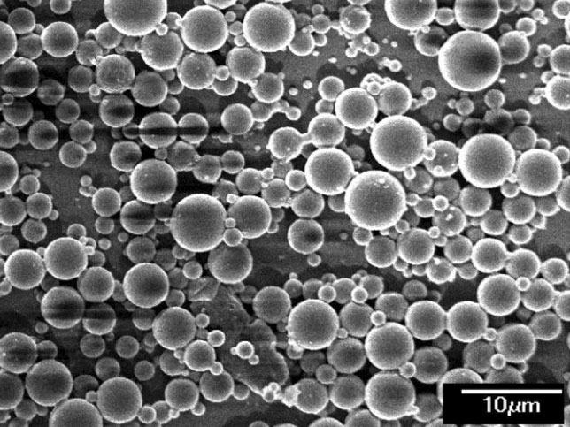
Lipid-coated PLGA
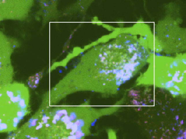
Gel particles in dendritic cells

Darrell Irvine is a Howard Hughes Medical Institute Investigator.  Our research is also supported by the National Institutes of Health, the Ragon Institute of MGH, MIT, and Harvard, the Bill and Melinda Gates Foundation, the Melanoma Research Alliance, the Bridge Project, and the Dept. of Defense (via the Institute for Soldier Nanotechnology and the Defense Advanced Research Projects Agency).  We gratefully acknowledge past support from the the National Science Foundation, the Arnold and Mabel Beckman Foundation, the Whitaker Foundation, the Human Frontiers Science Program, 3M, the Dept. of Defense Prostate Cancer Research Program, and the DuPont-MIT Alliance.